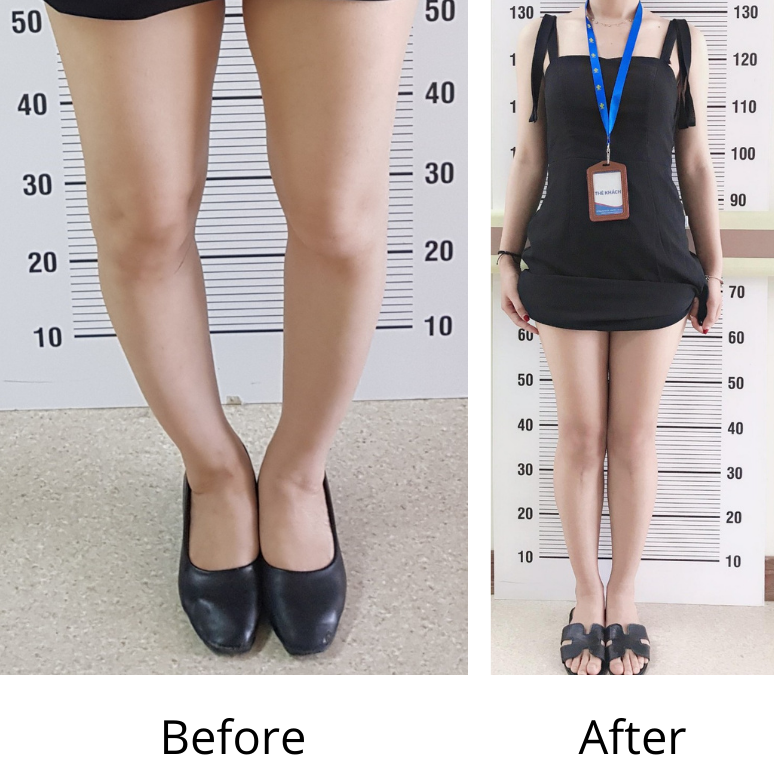
Bowlegs correction
Hyperextended elbow treatment
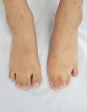
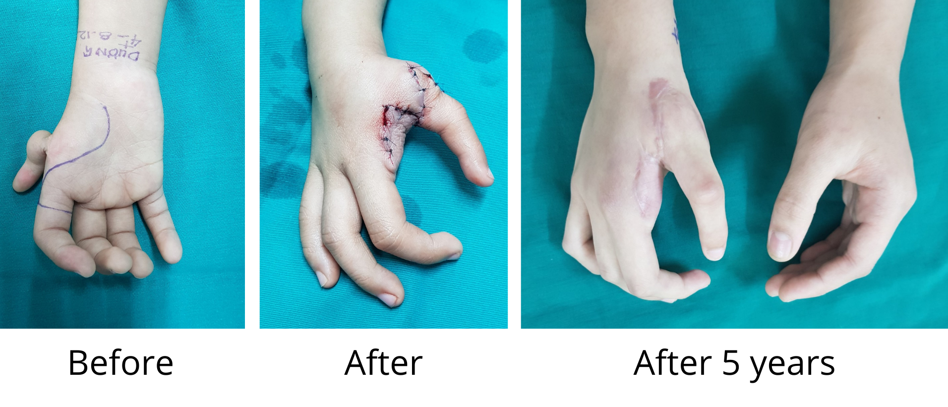
Toe deformities
Post-injury complications treatment through surgery
Bowlegs is not an uncommon condition. Bowlegs is mostly caused by birth defects, with a few cases were results of achondroplasia or osteosarcoma at the ends of long bones, especially the lower part of the thigh bone or the the upper part of the shinbone.
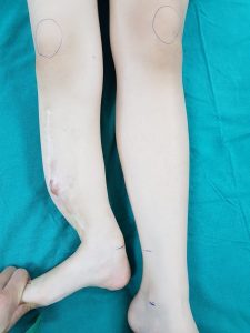
When there is deformity in the bones, the legs usually curve outward at the knees. Then the body weight is not evenly applied to both the medial tibial plateau and lateral tibial plateaus as usual, but applied mainly to the medial tibial plateau. Therefore, without treatment, it can lead to knee osteoarthritis. Usually after 45 years of age, knee joint pain begins to emerge. After 55 years of age, when the bones start to get osteoporosis, the deformity becomes more and more severe. Many cases have their knee joints replaced after 60 years of age. On the other hand, with bowlegs, the patient’s walk looks clumsy which can lead to lack of confidence, especially when the patient is wearing tight pants or short skirt. Therefore, patients with bowlegs should undergo surgery as soon as possible to avoid future complications and sequelae.
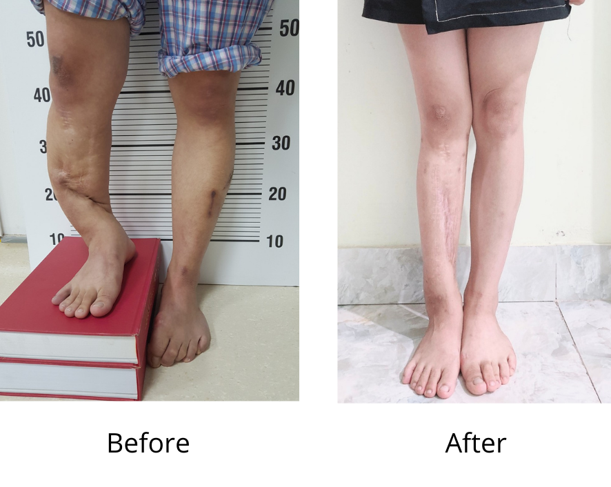
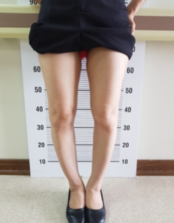
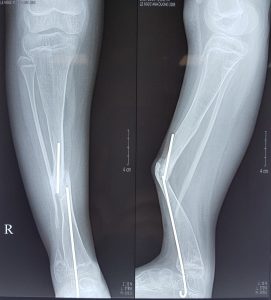
In order to determine if the patient has developed bowlegs, the simplest method is to examine the legs while the patient is standing. When the body weight is applied to both legs, the deformity can easily be detected. The examination should be done with the patient standing on a flat surface next to the wall. His heels, bottom, shoulders and head should touch the wall, both of the heels should touch each other and both of his feet should make a 60-degree angle. Normally in this stance, the two femoral condyles should touch or be 1-2cm from each other. When the two knees are 3-5cm apart, the patient’s bowlegs is still at an early stage. When they are 5-7cm, it is at a moderate level. When they are more than 7cm apart, the condition is severe. In order to accurately evaluate the axis of the leg bones, it is necessary to have an X-ray scan of the lower extremities, or a tomography to measure the angle between the tibia and femur. These measurements will help doctors accurately adjust during the surgery.
Regarding treatment, in young children, the majority of treatments are rehabilitation exercises and the use of orthopedic braces, but these treatments has produced limited results. In some cases with severe deformities, surgical intervention is required. The doctor would insert instruments inside to slow down the growth of the cartilage further and allow the bones to have a balanced growth. For people over 18 years old, surgical intervention is required to correct the bone axis.
Currently, there are many surgical methods for correcting bowlegs in adults, each has its own advantages and disadvantages. Dr. Doan has chosen the methods applied by famous surgeons in the world, which are axial osteotomy, bone grafting, compressing with locking splints. These methods have the following advantages:
When performing axial osteotomy, the surgical area should be just below the tibial plateau without any interference with the knee joints. This is the most curved bone, thus after the surgery, all deformities have basically been corrected (90-95% with other severe deformation). When performing axial osteotomy and grafting the bone into the cavity, the height of the body is increased by 1.5-2.5cm.
- The bone is compressed with a sturdy locking splints so no casting is required.
- After 2 weeks since the stitches are removed, the patient can start doing rehabilitation exercise.
- After 3 weeks, he can do walking exercises with crutches. After 6 weeks, he can walk normally.
- The incisions are small, only about 4cm, so the scars ensure aesthetics.
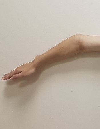
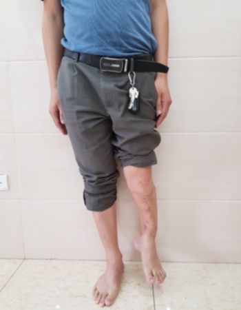
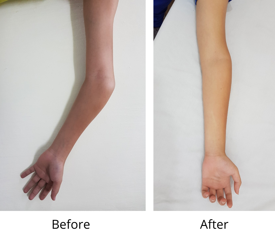
Hyperextended elbow is a common deformity, mostly caused by sequelae of the humerus condyle fracture since childhood. The humerus condyle is the most fragile part of the humerus, due to the thinness of the bones and development of the cartilages. Therefore, humerus condyle fracture is the most common type of bone fractures for children.
When there is a broken boke in this area, most of the treatments will include chiropractic and casting. Due to the complex nature of the anatomical structure, the straightening of all joints can seldom be performed. At the same time, because the fracture of the bones will affect the integrity of the growth cartilages, their nourishment after the fracture will also suffer severely. Therefore, when the bones are intact, their developments will cause the growths of the epiphyses to be uneven. Normally, the cartilages of the lateral condyles would have a ordinary growth. However, the cartilages of the medial condyles have a slow growth. Because of this, the bigger the body starts growing , the more severe the level of the hyperextended elbow gets.
In most cases, the elbow joint still functions normally. Movements like bending orstraightening the elbow, and turning of the forearm can be made normally. Despite the arm with the hyperextended elbow may feel weaker than the other arm, most patients decide to undergo surgery because of the cosmetic aspect of the deformity and the desire to get their arm straightened.
Surgery will only be indicated to patients at the age of 18 or above who are no longer in their physical development period. If the adjustment is made during the development period, there is a chance that the deformity wil reoccur. The only exceptions are cases of severe deformity with the open angle of more than 30 degrees. Regarding these cases, surgical procedure is allow for patients from 12 years old.
Regarding the techniques: The incisions are only 6-7cm. Behind the humerus, the doctor will perform osteotomy in the most curved area, then straighten the arm and reattach the bone with locking splints.
After 10 days since the stitches are removed, the patient can start doing exercise without the need of any casts. After 3 weeks, the patient can move as normal.
- Surgical treatments for crooked toes and fingers.
- Congenital malformation of the tibial prosthesis in children.
- Toe lengthening for patients with brachymetatarsia
- Separation of fingers and toes from deformities since birth or caused by burn scars.
- Thumb reconstruction surgery for patients with thumb hypoplasia.
- Reconstruction for burn sequelae, and injuries that cause finger and toe shrinkages.