Limb Lengthening Procedure
Pre-operative
Before deciding to perform the surgery, doctors need to carry out a clinical examination for the patient to learn about the patient’s medical history, including reproductive history, physical and stature development during adolescence, puberty, hormone levels, childhood illnesses, genetic diseases, etc. In addition, doctors will assess the patient’s psychological status, aspirations and desires.
After this, the patient will receive subclinical examination and specialized examinations to exclude skeletal and other systemic diseases with contraindications to limb lengthening. Patients are also consulted by the doctor, receiving clear explanation on the surgical procedure, hospital stay, treatment process during hospital stay and after discharge, duration of treatment, possible accidents and complications. Patients need to understand and be ready, including the process of post-operative exercising and rehabilitation.
During surgery
In all procedures, epidural analgesia will be used to ensure that for at least the first 3 days after surgery, there will be no pain. Patients can choose between endotracheal anesthesia, or spinal anesthesia with pre-anesthesia. The average operative duration is 2 hours.
Post-operative
During inpatient treatment
– Regarding medicines:
+ Antibiotics: The patient is given prophylactic antibiotics. Therefore, there will be 1 antibiotic injection 30 minutes before the surgery, followed by the use of injectable antibiotics in the next 24 hours after surgery. From day 2, switch to oral antibiotics for 5 days.
+ Analgesics: Perform multimodal analgesia. Before spinal anesthesia, the anesthesiologist placed an epidural analgesia line for continuous pain relief for the first 3 days after surgery. After 03 days, remove the analgesia line and switch to oral analgesics, combining 2-3 types of pain relievers. The oral doses will be gradually reduced until the patient is discharged.
+ Edema and hematoma medications: Will be used in oral form, or sublingual administration, for 05 – 07 days.
+ Venous thromboembolism medications: 01 tablet per day, use continuously until discharge.
– The nurse will change the wound dressing on the first day after surgery, then repeat it every 48 hours, wrappring sterile gauze to protect the nail.
– Apply cold compression, elevate the surgical legs: Cold compression has vasoconstrictive properties, therefore reduces swelling, edema, and pain; elevating the legs to reduce edema should also produce good pain reliever effect, the duration of cold compression and leg elevation should be 03 – 05 days. During this period, in order for blood to be circulated and avoid occlusion, it is recommended to do passive exercises or active movements gently for the knee and ankle joints; always place the leg fixators perpendicularly while at rest or sleeping to prevent future shortening of the Achilles tendons.
– After 5 days since the surgery, when the pain and edema are no more, the patient should do more active exercises, especially of the joints of the lower limbs, and stretch the heel tendons. When going to bed, always keep the pillow straight and do not sleep in a bent position.
– After 7 days since the surgery, the doctor will stretch the frames and instruct the patient to self-operate the external fixator frames to stretch the cavities at the rate of 1mm per day, divided equally into 3 times (the stakes on each extensor frame are already divided into 6 faces, turn a different face every 8 hours at the time marks of 6 am, 2 pm and 10 pm), turn clockwise, from left to right looking down from above.
– After 10 days since the surgery, remove stitches and take an X-ray of the legs for assessment, if the cavities are well stretched, the patient can be discharged from the hospital and receive outpatient treatment.
During outpatient treatment
– Change the bandages around the nails, wipe the dried scabs around the nails with physiological saline, then sterilize with 70-degree white alcohol or Betadine and wrap sterile gauzes around the nails once a day. If the nails cause inflammation or are oozing pus, take a picture and notify the doctor to decide if antibiotics should be given.
– Following the doctor’s instruction, the patient needs to stretch on his/her own. Note that even if the stretching process does not cause any pain, it is recommended that the patient should not turn the stakes more than 3 times a day to ensure the regeneration of bones, tendons, muscles, blood vessels and nerves. During the stretching process, if there is a lot of pain, the patient can adjust the stretching rate to twice a day, at 6-7am and 6-7pm. Do not turn the stakes more than 3 times a day just to remove the fixation more quickly, because that will not give the cavities enough time to develop, leading to poor development of the cavities and even the growth of the bones.
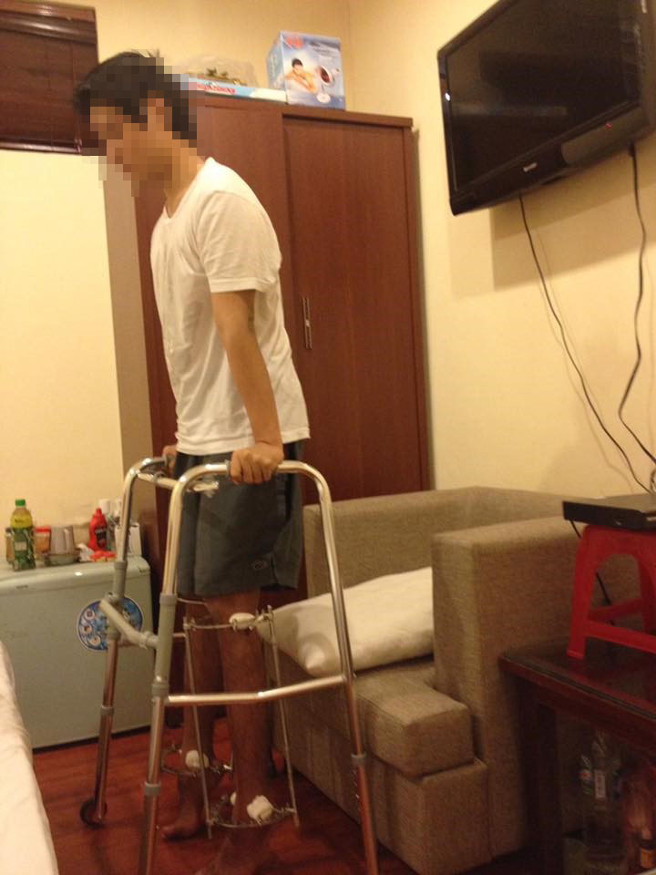
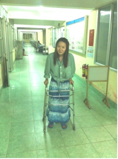
– Rehabilitation exercises during the process of stretching: Focus on exercises to stretch the knees, reduce hypertonia of the quadriceps, practice flexing the insteps into the shins to stretch the Achilles tendons, and practice rotation of the ankle joints. It is recommended to practice compressing part of your body weight when standing or walking with two crutches, or in a walker 3-4 times a day, 10-15 minutes each time. Note that while standing or walking, the body weight must be evenly distributed on 2 legs. Avoid putting the whole body weight on 1 leg.
– During the outpatient treatment, the patient needs to go to the hospital every 4 weeks to take X-rays and have clinical examinations; check if the screws holding the nails are loose, the nails are bent or the frames have skewed away; evaluate the development of the cavities to see if the growing speed is normal or not to decide whether to adjust the stretch speed, stop stretching, or schedule an appointment to remove the frames.
Treatment and exercises after the removal of the external fixator frame
Day 1: The patient receives intravenous fluids, analgesia, antibiotic injections and pain relievers. Leg is elevated.
Day 2: Change the first bandage, elevate the leg, have an X-Ray for examination after the removal of the fixator. During hospital stay, the patient should actively practice bending the knee and rotating the ankle joint to circulate blood and avoid muscle contraction.
After being discharged: Exercise to restore active and passive functions of the knee stretch, pratice stretching the Achilles tendon and improve ankle joint motion (practice by himself/herself or with the help of family members and friends).
Day 5: Pratice evenly applying a part of his weight to both legs while standing, so that the knees are straightened and the tendons of the heels are gradually stretched.
From day 7 to the end of the second month: When the nails are dry, start doing rehabilitation exercises (this is the best period to receive help from rehab technicians or to practice at rehab center), press for Achilles tendon to stretch, rotate the ankle in both clockwise and counterclockwise directions, practice knee extension, and do quadriceps strength training. When doing walking exercises, begin to walk with the help of a walker. After 2 weeks, cut 2 stitches where the bolts are screwed. Note that, at this stage, it is not possible to use armpit crutches or hand crutches yet because as the patient walks, the crutches will shift the body weight to one leg, which is not safe. The process of learning to walk will gradually increase the level of compression, which can be combined with indoor cycling to strengthen the quadriceps.
After 2 months: Come back for X-Ray for re-examination. The doctors then will assess the development of the cavities. Depending on natural disposition of the body and the degree of development of the cavities, the doctors will decide how long until the patient can switch to armpit crutches or hand crutches. If the cavities are well developed, the patient can switch to armpit crutches and allow full compression of the legs. At this stage, the patient should practice low-intensity exercises, indoor cycling, swimming, and wading.
After 4 months: Coming back for a check-up, the doctor will perform a minor surgery with anesthesia to remove the screws fixing the lower tibial joints (the patient does not need to be hospitalized) and assess the degree of osteotomies. If the cavities are good and the tendons are not tight, the patient can remove the crutches and do walking exercises, or walk with the help of hand crutches. This period encourages the practice of yoga and walking between 2-3 km per day.
From 6 months onwards: The patient can have an X-Ray and send it to the doctor for evaluation and instructions through phone. Normally, after 6 months since the removal of the fixators, the shin bones will be stable, the Achilles tendons will no longer be tight, the joints will be soft. The patient can walk as usual and can participate in moderate exercises, yoga, and swimming. Walking betweeen 4-5 km per day is recommended.
From 12 months onwards: When getting the X-Ray, if the doctors confirms that the bones are strong and firm, the patient will be able to perform high-intensity activities such as going to the gym, running and climbing.
After 24 months: When all sports activities can be performed as normal and the X-Ray shows healthy and strong bones, the patient can go and have a minor surgery to remove the intramedullary nails (can also be done after 3-5 years).
Illustration of rehabilitation exercises during outpatient treatment
- Applying a part of body weight to both legs while standing with external fixator frames
- Doing walking exercise with external fixator frames
Factors affecting bone healing and rehabilitation
In order for the bone healing process to be unevenful and rehabilitation process to be rapid, the following factors should be taken into account during leg lengthening process:
4.1. Factors related to techniques and practice
4.1.1. Stretching rate and pace
The optimal stretching rate for bone healing process is 1mm per day, divided into 3 times a day. A suitable stretching rate will ensure blood supply for bone tissues. The rate needs to be adjusted accordingly based on periodic clinical examinations and X-ray scans. If the bone formation is found to be poor, the stretching rate will be reduced or the stretching will be halted to improve bone formation. The halt can last from 3 to 10 days depending on age, gender, osteotomy position, bone quality, soft tissues status, etc. The waiting time will be long if poor bone formation progress is expected. The process will stretch nerves and blood vessel will also be stretched and grow to adapt to the maximum stretching rate of 1mm per day. However, during stretching, there can be numbness or pain in ankle and foot areas. Should these symptoms occur, the stretching pace can be reduced to twice a day. When it is reduced, the symptoms will subside and soon disappear after the stretching ends.
4.1.2. Exercising the joints and early compression
Exercising the joints and early compression help prevent muscular atrophy, joint stiffness as well as increase blood supply to the bone cavity. Many studies show that being compressed help create better bone cavity, increase bone density and produce more collagen I and II, BMP 2/4 and osteocalcin. The authors believe that early compression stimulates bone formation during limb lengthening.
Rehabilitation exercise also help slowly stretch the tendons, blood vessels and nerves in accordance with the stretching rate of the bone; avoid complications such as tendon shrinkage, numbness and pain during stretching; and improve leg function restoration after the fixator is removed.
4.2. Recommended medications and food during stretching
Be mindful that it is necessary for our foods and drinks to have enough nutrients. The meals should be varied. Avoid abstinence, especially from meat and fish to ensure the body absorb enough vitamins, minerals and protit, it is necessary to have an adequate diet of pork, chicken, beef, fish, seafood, etc. Avoid eating water spinach or drinking drinks made from water spinach as it will cause keloids.
4.2.1. Calcium-rich foods
- Cheese, yogurt
- Ocean fishes
- Flour, corn starch
- Amaranth, spinach
4.2.2. Functional foods
- Can take synthetic Vitamin (one tablet per day), or Vitamin 3B to enhance nerve regeneration (one tablet per day).
- Take Calcium – D3, one tablet per day.
Consultation
DIRECTLY
FROM DR. DOAN
Success stories